How To Read Nmr Results
NMR LipoProfile Test is FDA cleared for use in conjunction with other lipid measurements and clinical evaluation to aid in the management of lipoprotein disorders associated 7with CVD. The absorbance of energy to convert a nucleus from a 12 to a.
Nmr Spectroscopy
At minimum the spectral window should be 1 ppm to 9 ppm - for 1 H NMR and -10 ppm to 180 ppm for 13 C NMR.
How to read nmr results. Recall further that in the NMR experiment when and only when nuclei are irradiated with electromagnetic radiation of energy that exactly corresponds to the energy difference between the 12 and -12 spin states the nuclei absorb the energy and the NMR spectrometer measures this absorbance Review section 31 of the theory handout. Whether calculated or measured directly LDL-C is an. At other times however you will find that more data are necessary than solely a 1 H NMR spectrum.
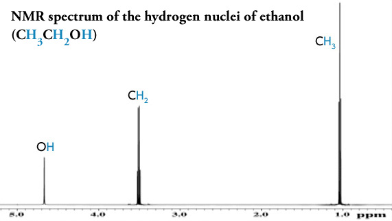
The analysis results were then compared with the l iteratures. In the presence of an external magnetic field B0 two spin states exist 12 and -12. And the peak at 26 is the methyl group which of course is joined.
Shows a method for getting all the useful information out of a proton NMR spectrum and using it to piece together the identity of an unknown molecule. Multiple landmark clinical studies show the higher the LP-IR Score the greater the risk of developing diabetes. Both VAP and NMR stuck 54 of the people into pattern B but tube gel electrophoresis classified just 5 two people as pattern B.
The resulting empirical formula is. Deciphering 1H NMR Spectra. The LP-IR Score is a weighted combination of six NMR lipoprotein variables that ranges from 0 most insulin sensitive to 100 most insulin resistant.
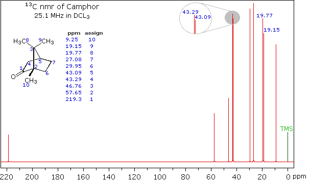
Combined analysis of 13 C NMR IR and other information may be needed for example. Double-bond equivalent also known as Degree of Unsaturation is calculated by a simple equation to estimate the number of the multiple bonds and rings. The two peaks at 137 ppm and 129 ppm are due to the carbons at either end of the carbon-carbon double bond.
Chemical shift values should be included. In the above case knowing the molecular formula conceiving of the possible. It assumes that oxygen O and sulfur S are ignored and halogen Cl Br and nitrogen is replaced by CH.
Df_NG_biomarker_metadata machine_readable_name colnames df_nmr_results NMR biomarkers here should be 250 stopifnot length nmr_biomarkers 250 Select only variables to be used for the model and collapse to a long data format df_long. As a result it is suggested to read more about the aromatics NMR in textbooks because they have got much more examples which will help you to learn how to interpret all those splittings. One of the most important concepts taught in organic chemistry is the method for determining the chemical structure of newly synthesized or unknown compounds.
Extract names of NMR biomarkers nmr_biomarkers. H NMR spectrum should be integrated. The following features lead to the nmr phenomenon.
The results varied considerably among the methods According to tube gel electrophoresis 79 of the people sampled fell into large fluffy pattern A LDL while VAP found that only 8 of samples were pattern A. A spinning charge generates a magnetic field as shown by the animation on the right. An unknown alcohol C5H10O has the following 1H NMR data.
25 The solvent peak should be clearly labeled on the spectrum. Looking for some more organic chemistry practice. The resulting spin-magnet has a magnetic moment proportional to the spin.
In this article we will summarize the concept of proton NMR the most common NMR information acquired by organic chemists. The step- by -step method on how to read the FTIR d ata was also p resented including reviewing simple to the complex. 2H doublet at 415 J 7Hz.
The 13 C NMR is a bit different from 1 H NMR in two aspects. NMR is a good technique for analyzing unknown compounds but it is limited in that it usually needs a bit larger amounts in microgram-scale at least and it should be 95 pure unless you want to. As you gain more skill at interpreting NMR data you may find that just a portion of the data is sufficient to determine a compounds identity.
H-NMR The first step in structural characterization is 1-dimensional proton H-NMR. Insets are encouraged to show expanded regions. 3H singlet at 163.
Use the coupon code Dave and save 20 right awayWhat are. 26 All peaks should be visible on the spectrum. Propose a reasonable structure.
The peak at just under 200 ppm is due to a carbon-oxygen double bond. Lipids Traditional lipid panel includes LDL-C HDL-C triglycerides and total cholesterol. 3H singlet at 170.
1H broad singlet at 383. 1 They are usually Decoupled therefore no splitting is seen in them. This post will walk you through the steps to fully characterize a molecule by 1- and 2-dimensional NMR including on how to perform NMR interpretation.

Proton Nmr Practice 1 Video Spectroscopy Khan Academy

Pin On Nmr Spectroscopy Practice Problems
![]()
Chemical Compound Proton Magnetic Resonance Spectroscopy Britannica

Integration In Nmr Spectroscopy Chemistry Steps
Nmr Basic Knowledge Nuclear Magnetic Resonance Spectrometer Nmr Products Jeol

Integration In Nmr Spectroscopy Chemistry Steps
6 6 H Nmr Spectra And Interpretation Part I Organic Chemistry

Nmr Spectroscopy Practice Problems

Pin On Nmr Spectroscopy Practice Problems
Deciphering 1h Nmr Spectra Organic Chemistry Help

5 3 Nmr Spectroscopy Chemistry Libretexts

5 3 Nmr Spectroscopy Chemistry Libretexts

What Compound Gives A Signal At 0 08 Ppm In The 1h Nmr Spectra Cd2cl2
.jpg)
Using Proton Nmr Spectroscopy For The Identification Of The Isomers Of Ethyl Acetate Butyric Acid And Isobutyric Acid

Pin On Nmr Spectroscopy Practice Problems

Organic Spectroscopy International Search Results For Vanillin Science Chemistry Organic Chemistry Chemistry Notes
Deciphering 1h Nmr Spectra Organic Chemistry Help

Pin On Nmr Spectroscopy Practice Problems

Pin On Orgo Cheat Sheets Tutorials And Reference Material
Post a Comment for "How To Read Nmr Results"